Short answer: yes.
Based on anatomy, brain imaging, and clinical research, the cymba conchae stands out as the most effective and reliable site for non-invasive vagus nerve stimulation (taVNS).
A unique gateway to the vagus nerve
Traditionally, activating the vagus nerve to influence inflammation has required surgical implantation of electrodes in the neck. While effective, this approach is invasive.
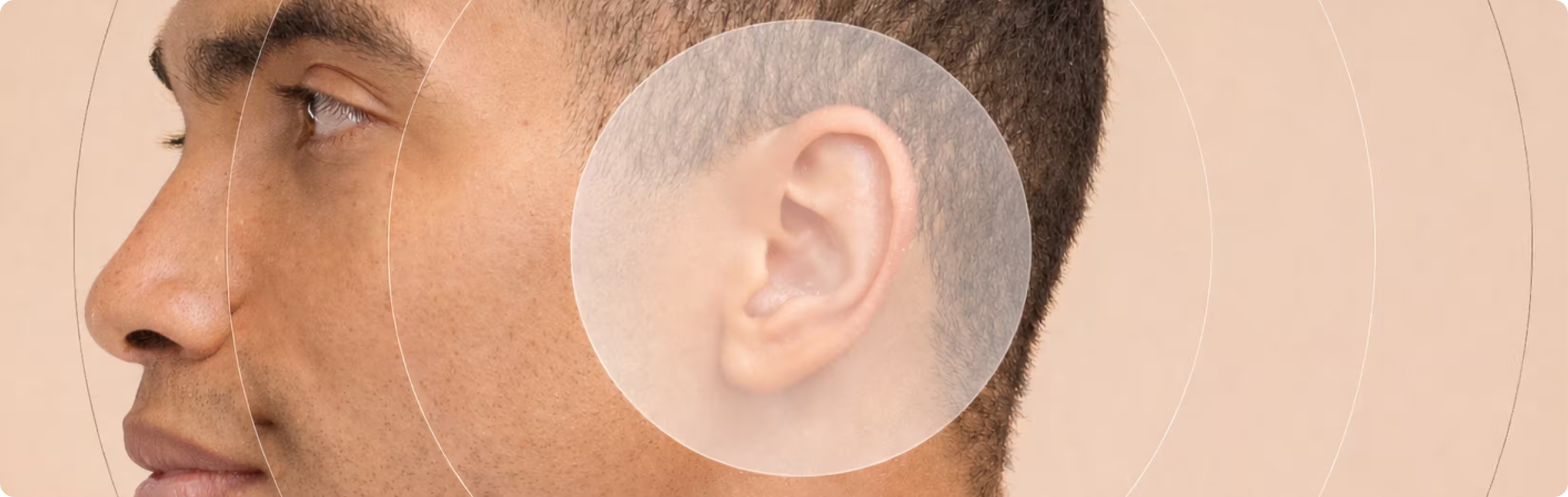
Transcutaneous auricular vagus nerve stimulation (taVNS) offers a non-invasive alternative by stimulating a small, specific area of the outer ear. This works because the outer ear contains the only place on the skin where sensory fibers of the vagus nerve are directly accessible.
Among all ear regions, the cymba conchae is unique:
- It contains superficial vagus nerve fibers
- The skin is thin, with minimal underlying fat
- Electrical signals can reach the nerve fibers efficiently
These features make the cymba conchae especially well-suited for precise, non-invasive stimulation.

What anatomy tells us
Not all parts of the ear are equal
Anatomical studies show that the auricular branch of the vagus nerve (ABVN) is the only branch of the vagus nerve that reaches the body surface.
While some vagal innervation may be found in parts of the ear canal or inner tragus, it is:
- Most consistently present in the concha
- Exclusively present in the cymba conchae
Importantly, cadaver studies demonstrate:
- 100% presence of vagus fibers in the cymba conchae
- Only ~45% presence in the tragus
This consistency matters. Reliable stimulation requires reliable anatomy.
What brain imaging shows
The cymba conchae activates the correct brain circuits
Functional MRI (fMRI) allows researchers to observe which brain regions become active during stimulation. Studies consistently show that stimulating the cymba conchae produces stronger and more specific activation of vagal pathways than stimulation of other ear regions.
Stimulation at this site activates key brainstem nuclei, including:
- The nucleus tractus solitarius (NTS)
- The locus coeruleus
These regions are primary entry points for vagal sensory signals and play a central role in initiating the cholinergic anti-inflammatory pathway.
Notably, the brain activation pattern seen with cymba conchae stimulation closely resembles that observed with invasive cervical vagus nerve stimulation—but without surgery.
Why this matters for inflammation and pain
To activate the inflammatory reflex, stimulation must engage ascending, brain-directed vagal signaling. Sensory vagus fibers are sufficient for this purpose—but only if they are reached reliably.
Because the cymba conchae:
- Is consistently innervated by the vagus nerve
- Provides direct access to sensory vagal fibers
- Produces robust activation of vagus-associated brainstem centers
…it is currently considered the optimal stimulation site for taVNS when the goal is to influence immune regulation and inflammation.