
This article explores how researchers study electrical stimulation of vagal pathways. It describes established anatomy and neuroscience while distinguishing mechanistic observations from evidence of specific clinical or wellness outcomes.
The nervous system communicates through electricity.
Every thought, movement and automatic bodily function depends on carefully coordinated electrical signals travelling through networks of nerve cells. The vagus nerve is part of this system, carrying information between the brain and many of the body's internal organs.
So what happens when an external electrical signal is applied to it?
The answer is more complex than simply "activating the vagus nerve."
Vagus nerve stimulation, or VNS, can influence electrical activity within vagal pathways. But the response depends on where stimulation is applied, which nerve fibers are engaged, how the electrical signal is delivered and which physiological pathways those fibers connect to.
Understanding these differences is central to modern research into VNS and bioelectronic medicine.
The vagus nerve is a two-way communication system
The vagus is the tenth cranial nerve and an important component of the parasympathetic nervous system.
There are two vagus nerves, one on each side of the body. They emerge from the brainstem and extend through the neck into the chest and abdomen, communicating with organs including the heart, lungs and gastrointestinal tract.
But the vagus is not simply a pathway through which the brain sends instructions to the body.
Most vagal fibers are afferent, meaning they carry sensory information from the body toward the brain. A smaller proportion are efferent, carrying signals from the brain toward peripheral organs.
The vagus should therefore be viewed as a bidirectional information network rather than simply an output pathway.
Sensory information travelling through the vagus reaches the brainstem, particularly an area called the nucleus tractus solitarius, or NTS. From there, information can be transmitted into wider neural networks involved in autonomic regulation and physiological homeostasis.1,2
This two-way anatomy is fundamental to understanding what happens during vagus nerve stimulation.
What does electrical stimulation actually do to a nerve?
Nerve cells communicate using electrical and chemical signals.
When a sufficiently controlled electrical pulse is applied to a nerve fiber, it can change the electrical potential across the nerve cell membrane. If the stimulus reaches the required threshold, an action potential can be generated.
That electrical signal can then travel along the nerve fiber.
VNS makes use of this basic property of nervous tissue.
Implanted vagus nerve stimulation has been studied for decades and is used clinically in specific medical contexts. Research on implanted cervical stimulation has reported measurable neural and physiological responses, but those findings are specific to the studied devices, parameters and populations.1
The scientific question is therefore no longer simply:
Can the vagus nerve respond to electrical stimulation?
It can.
The more interesting questions are:
- Which fibers are being stimulated?
- Where are they being stimulated?
- What electrical parameters are being used?
- Where do those signals travel?
- What happens when they reach the brain?
From implanted VNS to non-invasive stimulation
Traditional VNS involves surgically placing an electrode around one of the two cervical vagus nerves in the neck.
This provides direct access to the nerve and has generated decades of scientific and clinical knowledge.
Researchers subsequently began exploring whether vagal pathways could also be accessed without surgery.
Two principal non-invasive approaches have emerged.
Transcutaneous cervical VNS applies electrical stimulation through the skin of the neck in the region of the cervical vagus.3
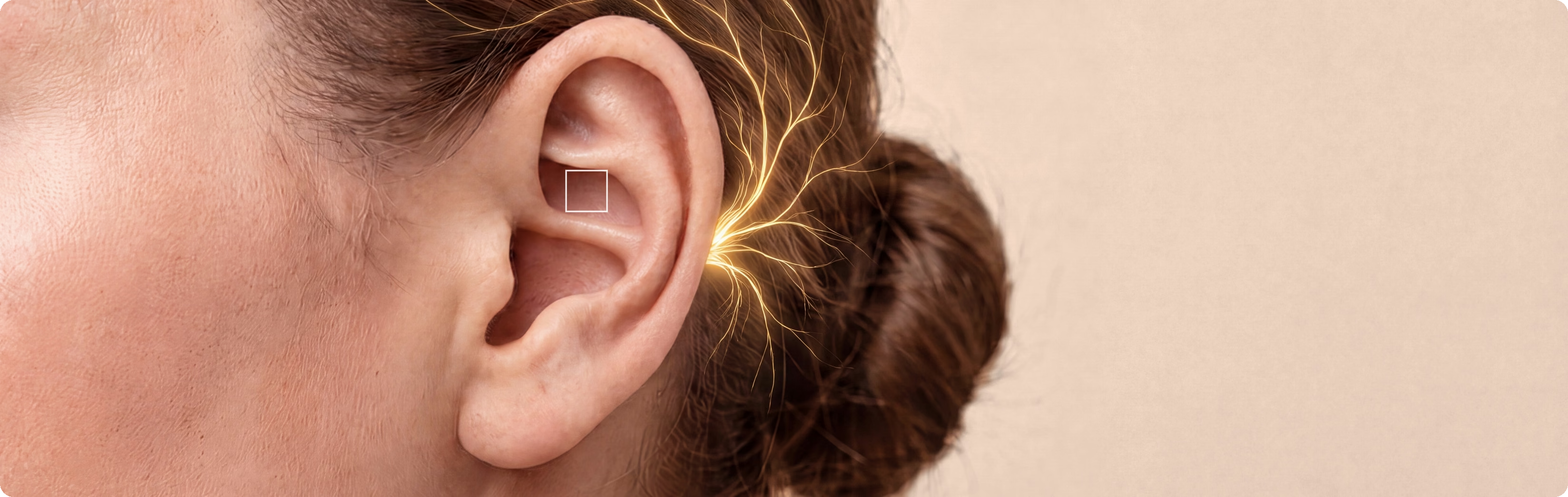
Transcutaneous auricular vagus nerve stimulation, or taVNS, applies stimulation to parts of the external ear innervated by the auricular branch of the vagus nerve.1–3
These approaches should not be considered interchangeable.
They stimulate different anatomical locations, and findings obtained with implanted cervical VNS cannot automatically be assumed to apply to auricular stimulation.
Together, however, they demonstrate that researchers can investigate vagal neuromodulation through several different anatomical approaches.
Why the ear provides an unusual window into the vagus nerve
The external ear has a complex sensory nerve supply.
It receives innervation from several nerves, including the trigeminal nerve, cervical spinal nerves and the auricular branch of the vagus nerve, or ABVN.
The ABVN is particularly interesting because it provides a location where sensory fibers associated with the vagus nerve reach an accessible skin surface.
Anatomical research has identified the cymba conchae, located in the upper part of the conchal bowl, as an area with substantial vagal sensory innervation.2,4
This anatomical observation provides the rationale for taVNS: electrical stimulation can be delivered through electrodes placed on the skin of the ear rather than through surgically implanted electrodes around the cervical vagus nerve.
This does not mean that every electrical stimulus applied to the ear selectively activates only vagal fibers.
Auricular innervation is complex. Individual anatomy, electrode placement, electrode design and stimulation parameters all matter.
For this reason, anatomical precision is an important consideration in taVNS research.
What happens in the brain during auricular stimulation?
One of the central questions in taVNS research is whether electrical stimulation of the ear actually influences brain regions associated with vagal sensory signaling.
Neuroimaging studies provide evidence that it can.
Functional magnetic resonance imaging, or fMRI, studies have reported changes in activity within brainstem and higher brain regions following stimulation of vagally innervated areas of the external ear.5,6
Of particular interest is the NTS, a major central destination for vagal afferent information.
Research comparing different auricular stimulation sites has reported measurable changes in the NTS and related brainstem activity during stimulation of the cymba conchae.6

From the brainstem, vagal sensory information connects with broader neural networks involved in autonomic regulation, arousal, stress responses and homeostasis.
These findings provide an important mechanistic bridge between the anatomy of the external ear and the wider physiological processes being investigated in taVNS research.
But there is an important distinction:
Evidence of neural engagement is not the same as evidence of a particular health outcome.
Changes observed on an fMRI scan demonstrate changes in neural activity. They do not, by themselves, establish that a specific clinical or wellness outcome will follow.
Vagus nerve stimulation and autonomic physiology
The autonomic nervous system continuously adjusts internal physiology without conscious effort.
Heart rate, vascular tone, digestion, respiration and many other processes are dynamically regulated through interactions between sympathetic and parasympathetic pathways.
The vagus nerve is an important component of the parasympathetic nervous system.
For this reason, researchers studying taVNS have examined physiological measures including:
- heart rate
- heart rate variability
- respiration
- sympathetic nerve activity
- other physiological biomarkers
Results are not uniform across all studies.
Differences in stimulation site, frequency, intensity, pulse width, session duration and experimental design make direct comparisons difficult.1,7,8
Heart rate variability, or HRV, is a good example.
HRV provides useful information about cardiovascular autonomic regulation, but it should not be interpreted as a simple measurement of "how active the vagus nerve is."
The vagus contains many different nerve fibers serving many different functions.
There is currently no single measurement that captures all of them.
The vagus nerve also communicates with the immune system
One of the most important advances in modern vagus nerve biology has been the recognition that the nervous and immune systems communicate extensively with one another.
Research beginning in the late 1990s identified neural mechanisms through which vagal signaling can influence inflammatory responses. This work contributed to the discovery of what became known as the inflammatory reflex.8,9
The principle extends beyond any particular disease.
The immune system does not operate independently of the nervous system.
Inflammatory signals arising in peripheral tissues can be detected by sensory pathways and communicated toward the brain. Neural signals can, in turn, interact with pathways involved in regulating immune-cell activity and inflammatory mediator production.
The vagus nerve participates in this bidirectional neuroimmune communication.
Research into these mechanisms helped establish an entirely new area of science: bioelectronic medicine, which investigates how precisely targeted neural signals interact with biological processes.
Experimental VNS has consequently become an important research tool for understanding how neural circuits interact with immune biology.
Clinical studies using implanted VNS have provided further evidence that electrically modifying vagal signaling can produce measurable biological effects in humans.
Importantly, these findings do not mean that all forms of VNS, all stimulation protocols or all non-invasive vagus nerve stimulation technologies produce equivalent effects.
Findings from implanted VNS or a particular experimental stimulation protocol cannot automatically be transferred to another VNS technology.
Each approach must be evaluated on its own terms.
Why stimulation parameters matter
Electrical stimulation is not simply "on" or "off."
A stimulation protocol is defined by several variables, including:
- stimulation location
- pulse frequency
- pulse width
- electrical intensity
- waveform
- electrode geometry
- electrode contact
- session duration
- repetition over time
Changing any one of these variables may change which nerve fibers are recruited and how neural circuits respond.
Two studies can therefore both describe themselves as investigating "taVNS" while using substantially different forms of stimulation.
This is one of the central challenges in interpreting the scientific literature.
Rather than asking simply:
"Does taVNS work?"
a scientifically more useful question is:
"Which stimulation parameters engage which neural pathways, and under what conditions?"
Can electrical stimulation have effects after stimulation stops?
The nervous system is not a static electrical circuit.
It can change its responsiveness based on previous activity, a property known as neuroplasticity.
Experimental VNS research has demonstrated that repeated stimulation, particularly when paired with specific activities or training, can be associated with longer-lasting changes in neural networks.10
VNS has therefore also been investigated as a method of pairing neural stimulation with specific experiences or rehabilitation training.
This illustrates an important principle of neuromodulation:
The duration of an electrical stimulus and the duration of the resulting neural response are not necessarily the same.
This does not mean that every form of VNS produces lasting effects.
How stimulation intensity, duration, repetition and timing shape neuroplastic responses remains an active area of research, and findings from specific experimental or clinical VNS protocols cannot automatically be generalized to other forms of stimulation.
Measuring vagus nerve stimulation is surprisingly difficult
If electrical stimulation is applied to a nerve, it might seem that scientists should simply be able to measure whether the nerve has been "activated."
In practice, it is much more complicated.
Researchers use several approaches, including:
- brain imaging
- electrophysiological measurements
- cardiovascular and autonomic measurements
- circulating biomarkers
- behavioral or physiological outcomes
Each provides a different window into the system.
None represents a universal measurement of vagus nerve activation.
This is particularly important when interpreting studies using HRV or other indirect biomarkers.
A change in one physiological measurement does not necessarily reveal which vagal fibers were stimulated or which neural circuits were responsible for the change.
Understanding VNS therefore requires converging evidence from anatomy, neuroscience, physiology and carefully controlled human research.
What does the science establish today?
Several conclusions can be drawn from decades of VNS research.
The vagus nerve responds to controlled electrical stimulation
Electrical stimulation can generate action potentials and neural activity within vagal pathways.1
Vagus-associated sensory pathways can be accessed through specific regions of the external ear
The auricular branch of the vagus nerve provides an anatomical route through which electrical stimulation can interact with vagal afferent pathways.2,4
Electrical stimulation of selected auricular locations can influence neural activity associated with vagal sensory processing
Human neuroimaging studies provide evidence of measurable changes in relevant brainstem and higher brain regions during auricular stimulation.5,6
The vagus participates in autonomic and neuroimmune physiology
The vagus is an important component of parasympathetic regulation and participates in bidirectional communication between the nervous and immune systems.8,9
How stimulation is delivered matters
Location, electrical parameters, electrode design and individual anatomy can influence which fibers are recruited and what responses are observed.1,3
And perhaps most importantly:
VNS is not one single biological intervention
Implanted cervical VNS, transcutaneous cervical VNS and auricular VNS involve different anatomical targets and stimulation approaches.
Findings from one technology, protocol or study population cannot automatically be generalized to another.
The next question is not simply whether VNS works
For many years, a central scientific question was:
Can electrical stimulation influence the vagus nerve?
Decades of neuroscience and clinical VNS research have established that controlled electrical stimulation can interact with vagal pathways and produce measurable neural and physiological responses.
The next questions are much more precise:
- Which fibers should be engaged?
- Where should stimulation occur?
- Which parameters produce reproducible neural responses?
- How should those responses be measured?
- How important is individual anatomical variation?
- And how do different patterns of neural engagement translate into different physiological responses?
Answering these questions is essential for understanding both the potential and the limitations of non-invasive vagus nerve stimulation.
It is also part of a broader scientific shift.
The brain, nervous system, immune system, cardiovascular system and internal organs are not isolated biological systems.
They continuously exchange information.
The vagus nerve is one of the pathways connecting them.
Electrical stimulation gives researchers a unique experimental tool: rather than introducing a molecule into the body, scientists can deliver a controlled electrical signal to a neural pathway and study what happens next.
That concept lies at the heart of bioelectronic medicine.